How I Took a MRI of a Very Small Part of my own Brain
I was the first person ever to record a fMRI signal from the thalamic reticular nucleus.
The TRN is small inhibitory structure in the brain that is implicated in attentional filtering. My boss at the time was the Director of York University’s MRI centre, Keith Schneider (and has since moved on to do the same and the University of Delaware), was mostly interested in how the subcortical regions of the brain contribute to attention: our proclivity to devote a lot of computational resources to only a few aspects of the data our brain could potentially access. Francis Crick (yeah, the DNA guy) was the first person suggest that the thalamus would implement these filters to perform attentional filtering, and this was confirmed many years later in experiments.
My job was to image this attentional filter in action by recording from the TRN in a person using functional MRI (fMRI). fMRI measures blood flow in the brain (the BOLD signal), in response to neural activity (when I was doing my degree, we still didn’t fully understand the relationship between neural activity and the BOLD signal, but now we much better understand it). The big problem with fMRI is that the signal to noise ratio is low, has a lot of geometric distortions as you crank up the resolution, and it is very susceptible to artifacts caused by movements. So we couldn’t just collect a brain scan, we had to collect a lot of very well-controlled, high resolution, noisy images and synthesize them into a functional picture of the TRN the same way astronomers take pictures of deep space.
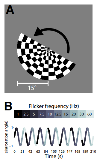
There were two big challenges: first, if we wanted to average many events together, we would need a really good idea of a particular event type that would drive TRN activity. We considered an attention switching task, but I found a lot of research that suggested different neural populations have different resonant frequencies, and that those resonant frequencies could be driven by external periodic stimuli, shown both in humans and animals.
To maximize the chance I would find the TRN, I didn’t want to rely on human behavior: I decided I just wanted to drive the human retina the same way animal researchers drop an electrode directly into the brain to zap it and record the response. I wanted a regular stimulus that could be down to people over and over again, that would give me a decent response. To do this, I started with something called “retinotopic mapping”. The primary visual system in the brain is organized like a reverse projector: different regions of the space in front of you map to specific locations on the cortex and thalamus. They call this the “retinotopic map”.
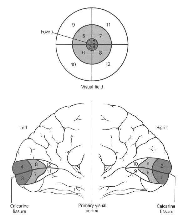
In fact, you can even project simple shapes onto the cortex and read them out using fMRI. A very expensive party trick. People map the “retinotopy” using a spinning disk at a fixed speed. Since they know the speed of the disk, they can look for brain regions that activate periodically at that frequency. My idea was to superimpose a flicker modulation on top of a spinning disk that stepped through frequencies at a different rate:
Any now for the coolest part: the retinotopic maps of my own lateral geniculate nucleus and thalamic reticular nucleus on top of a very high resolution brain scan:
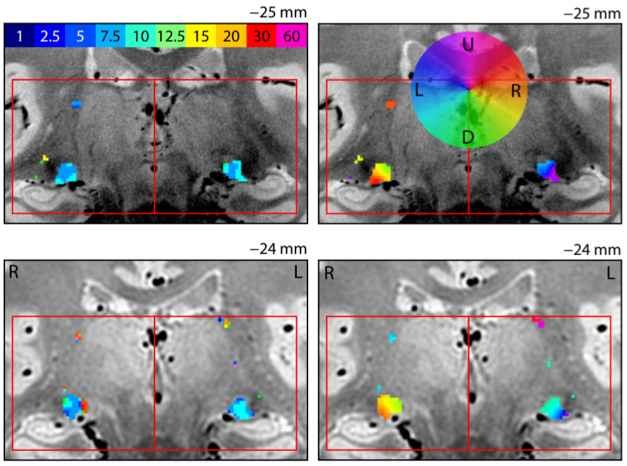
The main interesting thing we found was that the thalamic reticular nucleus seemed to be inhibiting the lateral geniculate nucleus at particular flicker frequencies: around 10-12 Hz, the lateral geniculate nucleus’s activity dominates (corresponding to the “alpha frequency” which is heavily implicated in visual attention), but interestingly, the thalamic reticular nucleus’ activity seemed to be maximal in the 20 - 60 Hz range where the lateral geniculate nucleus is less active: these frequencies correspond to the “beta/gamma frequencies” and are associated with top down feedback from non-visual areas (also see). It seems like driving your brain with a high flicker frequency sends shock-waves through your brain and activates an automatic feedback mechanism. I believe it: strobe lights are exciting at concerts for a reason.
This was my first big science project and I would have done a lot of things differently (including try to use an attention task to get a more interesting signal out of the brain). Aside from teaching me a tonne about physics and how the brain operates as distributed network, this was the project that taught me the basics of programming, and that kick-started my career.
If you want all of the dry details, here’s the paper.